 Section on Functional Imaging Methods
The Section on Functional Imaging Methods is within the Laboratory of Brain and Cognition and the National Institute of Mental Health. Functional MRI is a technique that utilizes time series collection of rapidly-obtained magnetic resonance images that are sensitive to localized brain activation induced hemodynamic changes. The utility of Functional MRI (fMRI) has been increasing since it was discovered in 1991. The limits of the technique (spatial and temporal resolution, interpretability of the signal, and applications) are determined by imaging technology, experimental and processing methodology, and the variable and incompletely-determined relationship between neuronal activity and hemodynamic changes.
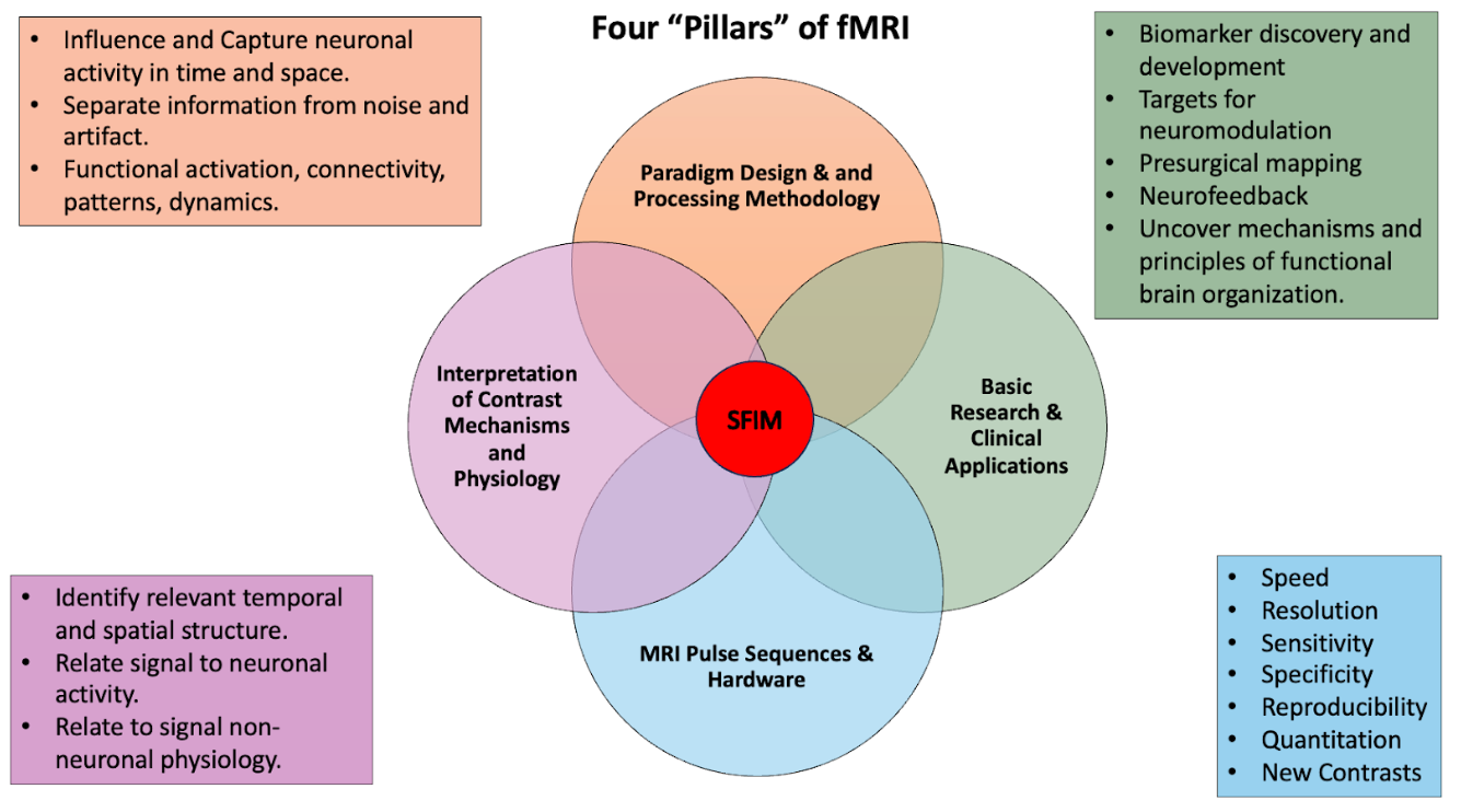
The work of SFIM is focused on pushing spatial and temporal resolution of fMRI as well as increasing its interpretability and ultimately the utility. This work roughly falls into four pillars:
